Home
/ Loculated Pleural Effusion Usg : Pleural Emphysema - Pleural effusion is a condition in which excess fluid builds around the lung.
Loculated Pleural Effusion Usg : Pleural Emphysema - Pleural effusion is a condition in which excess fluid builds around the lung.
Loculated Pleural Effusion Usg : Pleural Emphysema - Pleural effusion is a condition in which excess fluid builds around the lung.. Learn step 2 and shelf essentials in a free 10 min video. Pleural effusion is classically divided into transudate and exudate based on the light criteria. Inflammation of the pleura, called pleurisy. Pleural effusion is fluid buildup in the space between the layers of the pleura. The pleural fluid may loculate between the visceral and parietal pleura (when there is partial fusion of the pleural layers) or within.
More than one half of these massive pleural effusions are caused by malignancy; Pleural effusion is classically divided into transudate and exudate based on the light criteria. Excess fluid in the pleural space; Loculated effusions are collections of fluid trapped by pleural adhesions or within pulmonary fissures. Approximately 1 million people develop this abnormality each year in the united states.
Loculated Pleural Effusion Mesothelioma from lh5.googleusercontent.com Pleural effusions can loculate as a result of adhesions. Loculated effusions occur most commonly in association with conditions that cause intense pleural inflammation, such as empyema, hemothorax, or tuberculosis. Approximately 1 million people develop this abnormality each year in the united states. This situation most commonly is seen in patients with heart failure. Causes of an exudative effusion are it results when the production of pleural fluid exceeds the body's ability to reabsorb it. If none is present the fluid is virtually always a transudate. Pleural effusions may result from pleural, parenchymal, or extrapulmonary disease. Learn vocabulary, terms and more with flashcards, games and other study tools.
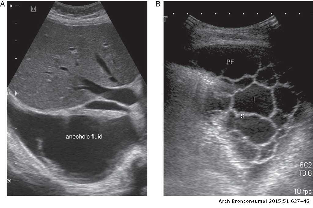
Obliteration of left costophrenic angle with a wide pleural based dome shaped opacity projecting into the lung noted tracking along the cp angle and lateral chest wall suggestive of loculated pleural effusion, however.
This situation most commonly is seen in patients with heart failure. Cancer, injury, or problems with other organs in your chest or abdomen, such as cirrhosis or pancreatitis. Inflammation of the pleura, called pleurisy. Pleural fluid/serum protein ratio >0.5. Reviewed by arefa cassoobhoy, md. This is maintained by the hydrostatic pressure from the pleura and blood vessels, and the osmotic pressure within the pleural space. Learn step 2 and shelf essentials in a free 10 min video. Send aspirated fluid for cytology. Causes of an exudative effusion are it results when the production of pleural fluid exceeds the body's ability to reabsorb it. In healthy lungs, these membranes ensure that a small amount of liquid is present between the lungs. Learn vocabulary, terms and more with flashcards, games and other study tools. The pleura are thin membranes that line the lungs and the inside of the chest cavity and act to lubricate and facilitate breathing. Occasionally, a focal intrafissural fluid collection may look like a lung mass.
Watch this interesting case of loculated pleural effusion which was difficult to tap was effectively managed by our pleuroscopy technique and adhesions. Learn about pleural effusion including causes of pleural effusion. Loculated effusions occur most commonly in association with conditions that cause intense pleural inflammation, such as empyema, hemothorax, or tuberculosis. Pleural fluid/serum protein ratio >0.5. Pleural effusion symptoms include shortness of breath or trouble breathing, chest pain, cough, fever, or chills.
Management of parapneumonic pleural effusion in adults ... from multimedia.elsevier.es Reviewed by arefa cassoobhoy, md. Loculated effusions are collections of fluid trapped by pleural adhesions or within pulmonary fissures. Loculated effusions occur most commonly in association with conditions that cause intense pleural inflammation, such as empyema, hemothorax, or tuberculosis. Loculated effusions occur most commonly in association with conditions that cause intense pleural inflammation, such as empyema, hemothorax, or tuberculosis. Differentiation of loculated effusions from solid masses. Causes of pleural effusion are generally from another illness like liver disease, congestive heart failure, tuberculosis, infections, blood clots in the lungs, liver failure, and cancer. Commonly from congestive heart failure or malignancy. e intrinsic characteristics of an effusion and its.
Reviewed by arefa cassoobhoy, md.
Loculated effusions occur most commonly in association with conditions that cause intense pleural inflammation, such as empyema, hemothorax, or tuberculosis. Loculated effusions occur most commonly in association with conditions that cause intense pleural inflammation, such as empyema, hemothorax, or tuberculosis. Approximately 1 million people develop this abnormality each year in the united states. Learn vocabulary, terms and more with flashcards, games and other study tools. Pleural fluid ldh > two thirds of upper limit for serum ldh. Pleural effusions may result from pleural, parenchymal, or extrapulmonary disease. They are encompassed within protective thin membranes called pleura. Accompanying adhesions can be identified. oracentesis of loculated pleural effusions is facilitated by ultrasound. Learn step 2 and shelf essentials in a free 10 min video. Most commonly caused by a viral infection. Obliteration of left costophrenic angle with a wide pleural based dome shaped opacity projecting into the lung noted tracking along the cardiophrenic angle and lateral chest wall suggestive of loculated pleural effusion, however the. Pleural effusion refers to a condition in which excessive amounts of fluid accumulates in the surrounding regions of the lungs.
If none is present the fluid is virtually always a transudate. Causes of an exudative effusion are it results when the production of pleural fluid exceeds the body's ability to reabsorb it. Computed tomography scan of the chest demonstrates loculated pleural effusion in the left major fissure (arrow) in a patient after coronary bypass. Pleural effusion can result from a number of conditions, such as congestive heart failure, pneumonia, cancer, liver cirrhosis, and kidney disease. This situation most commonly is seen in patients with heart failure.
Loculated Pleural Effusion / The Role Of Ultrasound In The ... from prod-images-static.radiopaedia.org Computed tomography scan of the chest demonstrates loculated pleural effusion in the left major fissure (arrow) in a patient after coronary bypass. Pleural fluid/serum ldh ratio >0.6. Pleural effusions occur as a result of increased fluid formation and/or reduced fluid resorption. Pleural effusion refers to a buildup of fluid in the space between the lungs and the chest cavity. Lung infections such as pneumonia or tuberculosis (tb). They are encompassed within protective thin membranes called pleura. This situation most commonly is seen in patients with heart failure. Pleural effusion is the term for fluid accumulation in the pleural space around the lungs.
This situation most commonly is seen in patients with heart failure.
Loculated effusions are mostly due to adhesions driven by pleural inflammation; Inflammation of the pleura, called pleurisy. Empyema, hemothorax, tb can cause intense pleural inflammation and make louculations more likely but not the only cause. Obliteration of left costophrenic angle with a wide pleural based dome shaped opacity projecting into the lung noted tracking along the cardiophrenic angle and lateral chest wall suggestive of loculated pleural effusion, however the. It has many causes (pneumonia, heart failure, blood clots, trauma. Differentiation of loculated effusions from solid masses. Pleura inflammation, causing sharp pain with breathing; If one of the following is present the fluid is virtually always an exudate. Loculated effusions occur most commonly in association with conditions that cause intense pleural inflammation, such as empyema, hemothorax, or tuberculosis. Learn vocabulary, terms and more with flashcards, games and other study tools. Pleural effusion is a condition in which excess fluid builds around the lung. If none is present the fluid is virtually always a transudate. Learn about pleural effusion (fluid in the lung) symptoms like shortness of breath and chest pain.
Occasionally, a focal intrafissural fluid collection may look like a lung mass loculated pleural effusion. Pleural effusions can loculate as a result of adhesions.